Th, 13.8.2026 | Original article from: CEITEC / Mgr. Halina Jílková
CEITEC MUNI researchers revealed unexpected gaps in the protein coat of tick-borne encephalitis virus, offering new insight into flavivirus assembly, maturation, and antibody neutralization.Tick-borne Encephalitis Virus Surprises Scientists: “Gaps” Discovered in Its Protein Coat
CEITEC/Plevka Lab: Tick-borne Encephalitis Virus Surprises Scientists: “Gaps” Discovered in Its Protein Coat
An international team led by researchers at CEITEC Masaryk University has, for the very first time, discovered that the tick-borne encephalitis virus is not as perfectly arranged as previously thought. Its surface contains areas where part of the protein coat is missing, leaving the viral membrane exposed. This incompleteness revealed that the rearrangement of surface proteins during viral maturation does not start simultaneously across the particle, but begins at a single point and then gradually spreads over the rest of the viral coat. The finding also suggests that the same principles apply to the entire group of flaviviruses, including dengue, Zika, and yellow fever viruses.
Using cryo-electron microscopy and electron tomography, the researchers observed thousands of tick-borne encephalitis virus particles and found that many deviate significantly from the idealized textbook model. “Flaviviruses are often depicted in textbooks as perfectly symmetrical particles. Our data, however, show that many immature and mature viral particles have exposed regions where part of the protein coat is missing. These imperfections helped us better understand how these viruses assemble and mature,” says Tibor Füzik from CEITEC Masaryk University (MUNI), lead investigator of the study. The arrangement of surface proteins at the edges of these exposed areas indicates that the coat of immature particles assembles step by step from individual proteins. New data further suggest that the rearrangement into a mature structure also occurs gradually, starting from a randomly formed center and expanding by adding individual protein units along its edges.
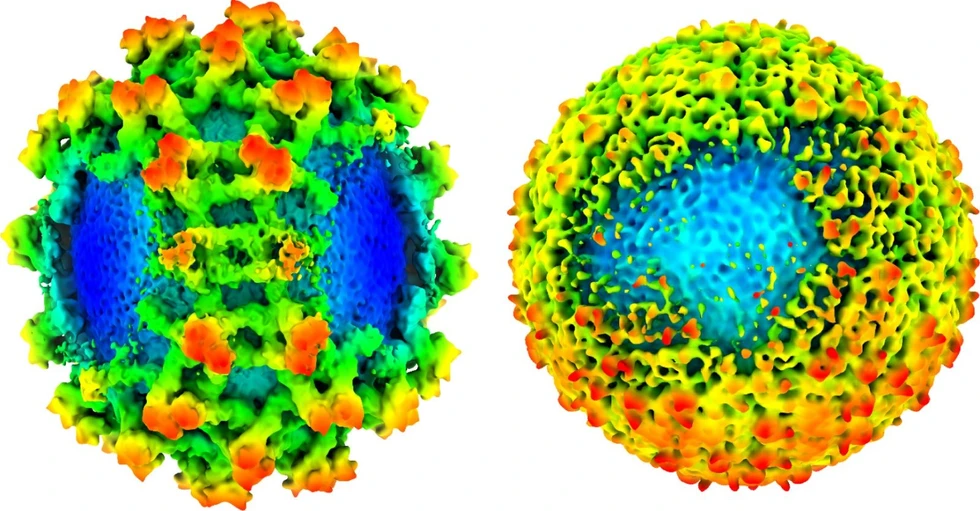
CEITEC/Plevka Lab: Tick-borne Encephalitis Virus Surprises Scientists: “Gaps” Discovered in Its Protein Coat: Immature (left) and mature (right) envelopes of tick-borne encephalitis virus particles contain disordered regions that expose the viral membrane.
Some parts of the viral surface proteins are normally hidden because they fit tightly together, making them difficult for antibodies to access. When part of the virus is not fully covered, these normally hidden protein regions are exposed, allowing antibodies – molecules used by the immune system to recognize and neutralize viruses – to attach. The discovery thus helps explain why some antibodies can effectively neutralize the virus, even though their target sites were previously thought to be inaccessible.
According to the study’s authors, this principle may not be limited to the tick-borne encephalitis virus. “We believe that similar rules of assembly and maturation may also apply to other flaviviruses, such as dengue, Zika, or yellow fever viruses, which cause millions of infections worldwide each year,” adds Pavel Plevka, head of the research group at CEITEC MUNI.
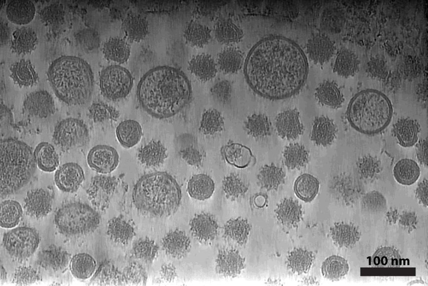 CEITEC/Plevka Lab: Tick-borne Encephalitis Virus Surprises Scientists: “Gaps” Discovered in Its Protein Coat: Tomographic analysis of tick-borne encephalitis virus particles shows irregularly shaped viral particles.
Thanks to these new insights into how flaviviruses assemble and mature into their infectious form, scientists can better understand how the immune system recognizes viral particles and use this knowledge to inform the future development of antivirals or vaccines.
The research was conducted in collaboration with scientific teams from the University of Helsinki and the Faculty of Science at Masaryk University.
The study was published in Science Advances.
The original article
Structures of asymmetric particles of tick-borne encephalitis virus provide insight into flavivirus assembly and maturation
Tibor Füzik, Maria Anastasina, Peter Pajtinka, Ausra Domanska, Lauri L. A. Pulkkinen, Lenka Smerdová, Lucie Nepovímová, Petra Formanová-Pokorná, Petra Straková, Jiří Nováček, Daniel Růžek, Robert Vácha, Sarah J. Butcher, Pavel Plevka
Sci. Adv., 2026, 12, eaee4765
https://doi.org/10.1126/sciadv.aee4765
licensed under CC-BY 4.0
Abstract
Flaviviruses are globally distributed human pathogens. However, the mechanisms underlying flavivirus assembly and maturation remain poorly understood. Here, we show that many particles of tick-borne encephalitis virus (TBEV) are asymmetric and lack subsets of surface heterodimers. Immature particles of TBEV contain incomplete spikes, providing evidence that their coats assemble directly from heterodimers of premembrane (prM) and envelope (E) proteins. Exposure of TBEV particles to acidic pH in the Golgi complex promotes maturation. The spikes and herringbone regions in TBEV maturation intermediates are oriented randomly rather than conforming to a common icosahedral symmetry. Consequently, the mature herringbone lattice forms around a randomly oriented nucleation center, expanding by addition of membrane-envelope heterodimers as the spikes disassemble and prMs are cleaved. The observed incompleteness of the protein coats explains, as an alternative to particle breathing, how flaviviruses can be neutralized by antibodies that bind to regions of E proteins normally inaccessible in the spiky or herringbone structures.